A burial in southern Peru has given researchers an unusual glimpse into a rare congenital condition that affected a child around 1,600 years ago. The infant, buried in a cemetery belonging to the ancient Nasca culture, had cyclopia, a severe developmental condition in which the eyes fail to form separately, resulting in a single eye socket in the centre of the face.An analysis of the child’s skeletal remains and ancient DNA has linked the condition to trisomy 13, in which a person has an extra copy of chromosome 13. The findings, published online in the journal Proceedings of the National Academy of Sciences (PNAS) on November 9, 2026, combine archaeological, skeletal and genetic evidence to investigate the condition and the circumstances of the burial.
What researchers found in the 1,600-year-old burial
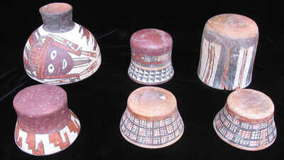
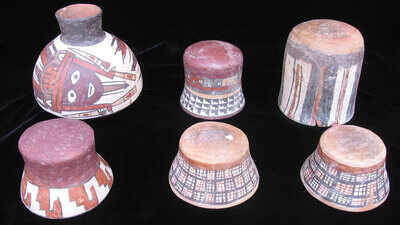
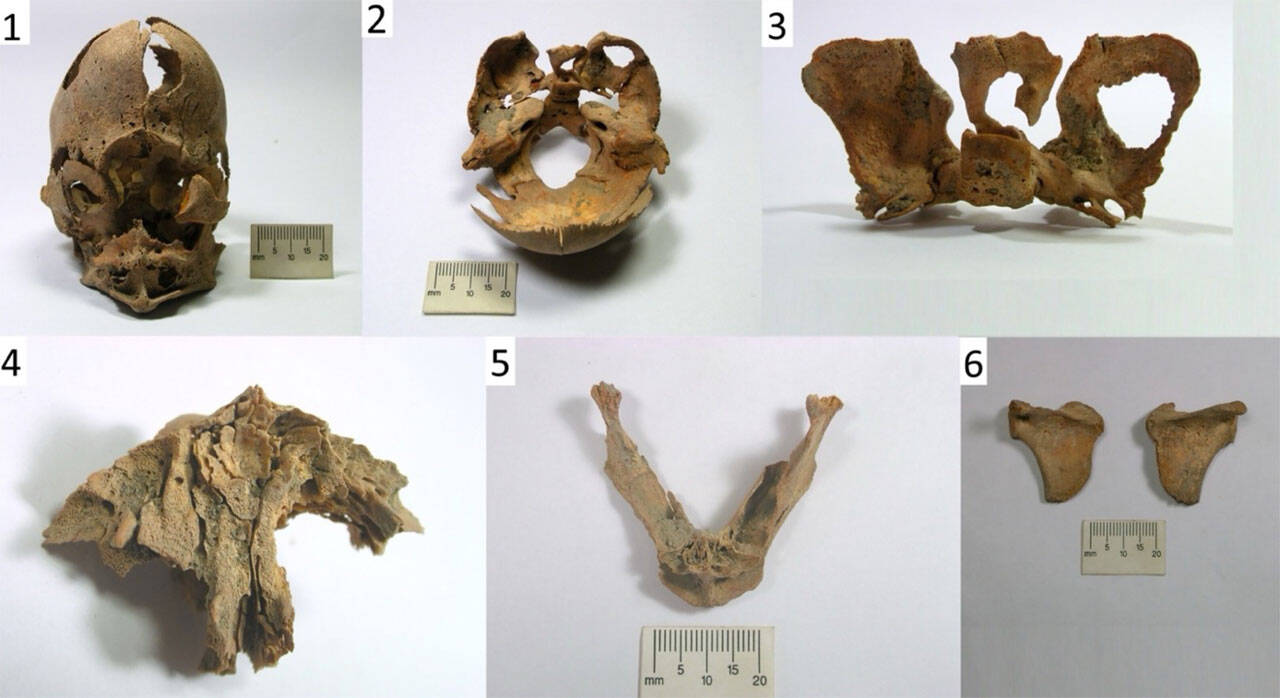
The remains were recovered from Burial 25 at Jauranga, an archaeological site in the Palpa Valley of southern Peru. The burial dates to the Middle Nasca period, between 325 and 440 CE.Researchers examined the remains of two infants placed one above the other inside a ceramic vessel. One, identified as JA13A, had several unusual craniofacial features, while the other, JA13B, showed no comparable congenital skeletal variations.According to the PNAS study, JA13A’s skull had a single median orbital cavity, meaning there was one eye socket positioned centrally on the face. The remains also showed an absence or severe underdevelopment of several midline facial structures, including the nasal bones and nasal cavities. Other skeletal differences included an unusually shaped skull, a narrowed left clavicle, abnormal curvature of the shoulder blade and two malformed vertebrae.The researchers found that the skull’s features matched clinical descriptions of cyclopia, the most severe craniofacial manifestation of a condition called alobar holoprosencephaly. This develops when the embryonic forebrain does not separate completely during early development.
A 1,600-year-old infant burial in Peru reveals a rare genetic condition; ancient DNA links cyclopia to trisomy 13. (Photo: pnas.org)
Photographic views of selected cranial and postcranial elements showing the morphological traits observed in individual JA13A: 1) articulated skull, frontal view; 2) internal view of the skull base; 3) sphenoid, posterior view; 4) maxilla, palatal view; 5) mandible, superior view; 6) scapulae, posterior view, showing a notch below the spine on the medial border. The cranial bones in panels 1 to 3 were temporarily articulated with removable adhesive for presentation purposes. (Photo: pnas.org)
Ancient DNA identifies an extra chromosome 13
To investigate the possible genetic cause, the researchers analysed ancient DNA recovered from the infants’ skeletal remains. Their findings showed that JA13A had an additional copy of chromosome 13, a condition known as trisomy 13.The team used three independent analytical approaches to examine chromosome numbers and compared the results with data from 250 ancient South American genomes. All three methods detected an excess of chromosome 13 material in JA13A, supporting the diagnosis of trisomy 13.The study identifies this chromosomal abnormality as the likely cause of the infant’s cyclopia. Trisomy 13 is a recognised genetic cause of severe holoprosencephaly, although the condition can have other causes as well.The researchers also established that both infants were genetically female. However, their mitochondrial DNA showed different maternal lineages, indicating that they were not maternally related.
Could the infant have survived after birth?
Cyclopia is extremely rare, occurring in approximately one in 100,000 births, and is typically fatal before or shortly after birth.The researchers found an interesting difference in the developmental indicators from JA13A’s remains. Dental development suggested an age of around two to four months after birth, while measurements of the long bones corresponded to late fetal or perinatal development.This could indicate that the infant survived beyond the immediate period around birth. However, the researchers cautioned that the condition and trisomy 13 may have affected normal dental and skeletal growth. The available evidence therefore cannot establish the child’s exact age or confirm how long the infant lived after birth.The paper, led by Elsa Tomasto-Cagigao and colleagues, also stresses that the remains cannot establish whether the child received care or how the condition was understood by the community.
What the burial reveals about Nasca funerary practices
The two infants were placed together inside a ceramic vessel, a form of burial documented among Nasca communities. According to the researchers, urn burials accounted for around one-quarter of documented Nasca graves in the Palpa region and were used predominantly for children younger than six years.The burial contained no additional grave goods. Its treatment was not archaeologically distinct from other documented Nasca child burials in the region.The researchers suggest that the child was incorporated into an established funerary tradition despite the unusual physical condition. However, they caution against drawing conclusions about the infant’s social identity, treatment or care during life from the burial alone.The study also discusses other archaeological evidence from the Andes showing varied historical responses to bodily differences, including examples of care, social inclusion and exclusion in different communities and periods. These examples cannot be treated as direct evidence of how the Middle Nasca community viewed this particular child.By bringing together ancient DNA, skeletal analysis and archaeological evidence, the research provides a rare account of a congenital condition in a prehistoric individual. It also demonstrates how genetic analysis can help researchers investigate the biological origins of conditions identified in ancient human remains.